Enjoy the event with subtitles available in multiple languages. *Subtitles are AI-generated and may contain inaccuracies.
close
This content is not available in your location
This event is scheduled for
Only logged in users can watch the content
To access AIS Channel content, please allow all cookies. Please click here to configure your preferences.
The Chinese Perspective: Uniportal VATS Right Upper Lobectomy
Johnson & Johnson MedTech
Share
share
Jul, 2022
Chat
keyboard_arrow_down
Description
keyboard_arrow_down
In recent years, China's thoracic surgery technology has developed rapidly,
entering the era of minimal invasive precision and committed to promoting
the standardization and precision of thoracic surgery. AIS specially invites
the leading faculties in the field of thoracic surgery from China, Italy and
the United States to share their academic expertise on the Uniportal VATS
Right Upper Lobectomy surgery.
Focusing on the operation, technical difficulties and innovation, AIS is
offering a rich and diversified academic feast in the form of an explanatory
operation video, case sharing and interactive discussion through the
Telesurgeon Technology. Through a systematic approach, we can further
explore surgical skills and learn difficult techniques, continuously improving
the technical standards of thoracoscopic lobectomy in the world, so that
the vast majority of patients can benefit.
PROGRAM
Welcome and Introduction – Dr. Ke En Oh (China)
Case Introduction – Dr. Jiao Heng (China)
Right Upper Lobectomy. Part 1: The incision Position – Dr. Tan Lijie (China)
Key Decision 1 Question – Dr. Marco Scarci (Italy) and Dr. Tan Lijie (China)
Right Upper Lobectomy. Part 2: The Anterior Approach – Dr. Tan Lijie (China)
Key Decision 2 Question – Dr. Michael Zervos (USA) and Dr. Tan Lijie (China)
Right Upper Lobectomy. Part 3: Lymph Nodes Dissection (Station 10, 4R, 2R & 7) – Dr. Tan Lijie (China)
Key Decision 3 Question – Dr. Marco Scarci (Italy) and Dr. Tan Lijie (China)
Right Upper Lobectomy. Part 4: Suture of Incision & Fixation of Chest Tube – Dr. Tan Lijie (China)
Key Decision 4 Question – Dr. Michael Zervos (USA) and Dr. Tan Lijie (China)
Discussion and closing remarks – Faculty moderated by Dr. Tan Lijie (China
CASE
Clinical History:
A 41-year-old male patient diagnosed with a lesion in the right upper lobe 3 weeks ago. No signs of Pulmonary Embolism.
Preoperative Examinations:
Pulmonary function:
FEV1:2.21L
FEV1%: 105 of predicted value
EKG:(-)
Abdominal and cervical ultrasound: (-)
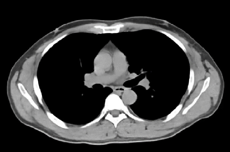
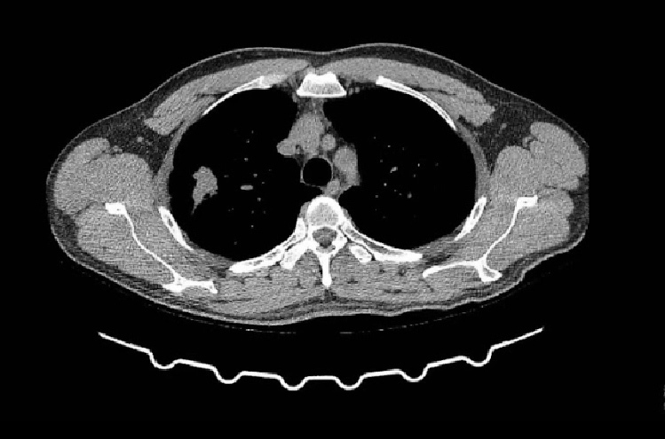
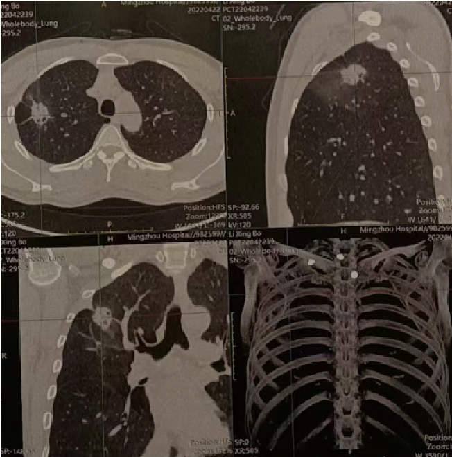
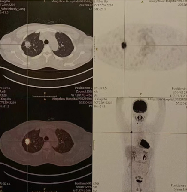
CT scan:
a solid lesion in right upper lobe
Size: 2.8X 2.3cm
Lobulation, burr and local traction of adjacent pleura can be seen at the edge
No hilar and mediastinal lymphadenopathy
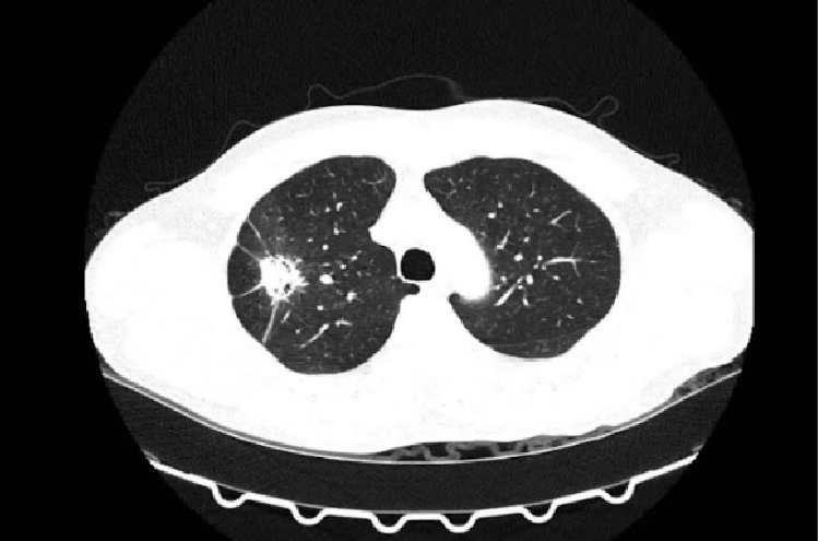
PET-CT:
A solid lesion in right upper lobe
Size: 2.3X3.0cm;SUVmax: 13.9
Also no hilar and mediastinal lymphadenopathy
Diagnosis and surgical strategy:
Right upper lobe lesion, highly suspicious of NSCLC
cT1cN0M0, stage Ⅰ A3
VATS RUL lobectomy
System lymph node dissection
Pathology:
pT1cN1M0, stage IIB
Invasive adenocarcinoma, grade III, alveolar and papillary(80%), micropapillary(20%)
Station 11s:2/4; 2R:0/7; 4R:0/5; 7:0/4; 10R:0/3;
Mutation of epithelial growth factor receptor (EGFR) exon 21(L858R)
Department introduction
35 staff surgeons, divided into 6 groups with 129 beds. more than 9000 thoracic surgeries annually.
Faculties
Dr. Qun Wang M.D. FRCS Chief Physician
Dr. Lijie Tan M.D. FACS Chief Physician
Dr. Di Ge M.D. FCCPChief Physician
Procedure trained
- Uniportal Vats Lobetomy
- Precise Location % Segmentoctomy for Pulmonary Nodules
Clinical Solution
- (Pre-OP) Patient indication, OR set up,
- (In-OP) Dissection technique of vessels, lobes resection. Avoiding complication
- (Post-OP) Managing complication
Technical Solution (safety use)
Harmonic 1000i, Powered Echelon flex+, (GST, PVS)
Learning Objectives
This course will help the participants to master the Uniportal Vats technique more proficiently
Target customer / Criteria
Fully Qualified physicians who already have experience in VATS and going to move to uniporta VATS.
Highlight for this course Uniportal VATS - Lobectomy Majority
- The World's First Book of Uniportal VATS
- Uniportal Video-assisted Thoracic Surgery by Dr. Lijie Tan
VATS Segmentectomy - Hi-End VATS Segmentectomy
- Precise Location & Segmentectomy for Pulmonary Nodules
Yet, the VATS segmental surgery is
increasing both in quantity and
quality. We use pre-op 3D model to
within the lung parenchyma.
Thank you for your interest! You will be contacted by the Shanghai Zhongshan Hospital


 Dr. Qun Wang M.D. FRCS
 Dr. Lijie Tan M.D. FACS
 Dr. Di Ge M.D. FCCP
Chief Physician
Dr. Di Ge M.D. FCCP
Chief Physician


