search
A 83-year-old female with a previous history of bronchial asthma. The patient arrived at the emergency room due to 6 days’ constipation and diffuse abdominal pain, with no nausea or vomiting and no fever.
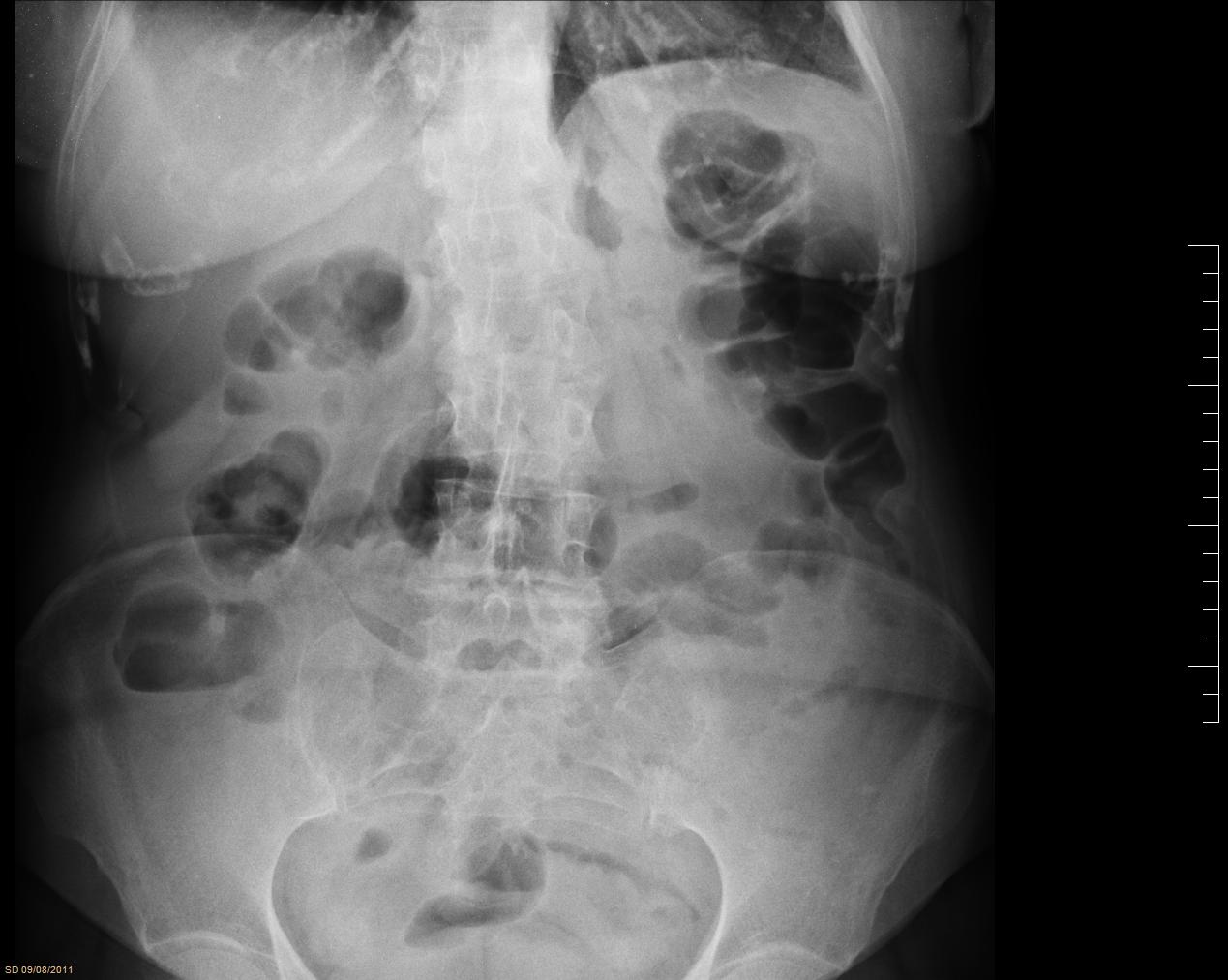
Vital signs were stable, the abdomen was soft with pain on deep palpation diffusely. DRE was normal, with no stool in the rectal vault. The blood test came back with leukocytosis and elevated CRP and the X-ray was normal (FIG. 1). It was decided to request a CT-Scan by the symptoms and the analytical results.
An abdominal CT-Scan with oral and intravenous contrast revealed, at the level of the left iliac fossa, a linear hyperdense image inside a loop of bowel (proximal ileum) and trabeculae adjacent mesenteric fat. These findings supported the diagnosis of a foreign body stuck in the intestine (VID. 1) with inflammatory changes, no pneumoperitoneum and no free intra-abdominal fluid.
An exploratory laparoscopy was carried with the patient in the supine position with open legs, with the leading surgeon and its assistant on the right of the patient. A total of 3 trocars were used: a 12mm trocar in a umbilical position for a 30º scope, a 12-mm trocar at the right iliac fossa and a 5mm trocar at the right flank.
The hyperemic small bowel was objectified and huddled together on the sigma. There were no other findings in the abdominal cavity. We decided to perform a Pfannenstiel assistance incision to explore the bowel affected. A 3M protective bag was placed on the incision, the intestine was extracted, and we objectified a punctate perforation secondary to a foreign body presented here (bone meat). The small intestine had unfavorable local characteristics at the perforated zone so we proceeded to perform resection of the affected bowel and a T-T manual anastomosis.
The surgery took 75 minutes and was uneventful. The patient started oral intake on the first postoperative day and left hospital three days after surgery. Histopathology revealed a traumatic intestinal perforation with associated peritonitis. In outpatient follow-up, the evolution was good, with no incidents.