search

A 82 years female, with overweight (BMI of 31 Kg./m2) patient with previous history of high blood pressure presented to the emergency department complaining of abdominal pain and distention. At physical examination there was tenderness at the left iliac fossa and a X ray revealed colonic distention.
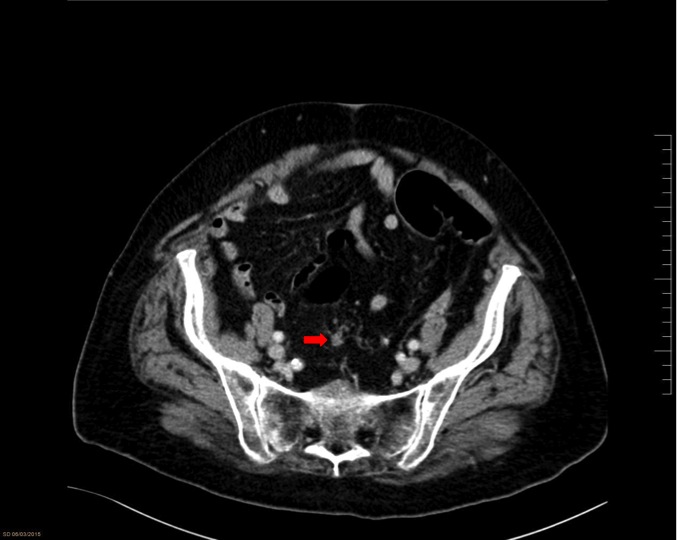
The patient was admitted into the hospital for further investigation. She remained stable without criteria of sepsis or complete bowel occlusion. A CT Scan revealed pathological lymph nodes at the pelvis (FIG. 1), the sigmoid colon seemed to have irregular walls (FIG. 2).
In order to achieve a more precise diagnosis the radiologist decided to administer contrast by enema, this strategy revealed a stenosis of the sigmoid colon (VID. 1) and led her to give the preliminary diagnosis of colonic carcinoma.
A colonoscopy showed a circumferential lesion at 30cm from the anal verge, the biopsy was inconclusive.
Nutritional support was administered until normalization of the pre-albumin levels. Elective sigmoidectomy with primary anastomosis by laparoscopic approach was carried out without incidences.
She had a correct post-operative course and was discharged the 3rd day after the procedure. Pathological examination was compatible with a pT3N1 moderately differentiated adenocarcinoma.